OBSERVING THE NAIL SHAPE AND SURFACE
|
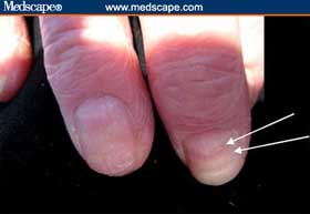
Clubbed Fingernails:
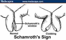
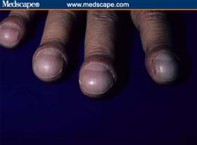
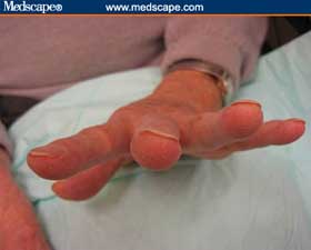
Nail clubbing involves a softening of the nail bed with the loss of normal Lovibond angle between the nail bed and the fold, an increase in the nail fold convexity, and a thickening of the end of the finger so it resembles a drumstick. To determine whether nails are clubbed, have the patient place both forefinger nails together and look between them. If you can see a small diamond space between them (Schamroth's window) then the nails are not clubbed (Schamroth's sign) (Figure 2). Causes of clubbing (not exhaustive) include the following (Figure 3): |
|
|
Lung cancer, pulmonic abscess, interstitial pulmonary fibrosis, sarcoidosis, beryllium poisoning, pulmonary arteriovenous fistula, subacute bacterial endocarditis, infected arterial grafts, aortic aneurysm Inflammatory bowel disease, sprue, neoplasms (esophagus, liver, bowel) Note: Chronic obstructive pulmonary disease does not cause clubbing. |
|
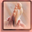
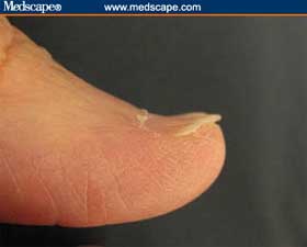
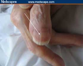
Koilonychia:
Koilonychia are spoon-shaped concave nails (Figures 4). This occurs normally in children and usually resolves with aging. To determine whether a nail is spooned, perform the water drop test. Place a drop of water on the nail. If the drop does not slide off, then the nail is flattened from early spooning. An experienced clinician can look at the nail and perform a "mental" water drop test. Causes include the following: |
|
|
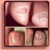
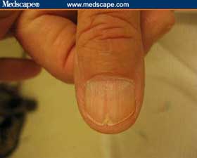
Beau Lines:
In 1846, Joseph Honoré Simon Beau described transverse lines in the substance of the nail as signs of previous acute illness. The lines look as if a little furrow had been plowed across the nail. Illnesses producing Beau's lines include the following: |
|
Intermittent doses of immunosuppressive therapy or chemotherapy can also produce Beau's lines. Severe zinc deficiency has also been proposed as a cause of Beau's lines. By noting its location on the nail, the approximate date of the illness associated with it can be determined (Figures 5A, 5B). Moreover, the depth of the line provides a clue to the severity of the illness. |
|
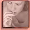
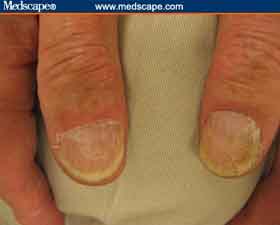
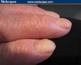
Thin Brittle Nails:
Thin, brittle nails can indicate the following (Figure 6): Figure 6: Note the thin nails in this woman with severe osteopenia. |
|
|
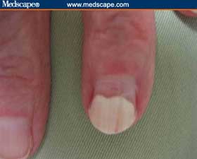
Onychorrhexis:
Onychorrhexis is the presence of longitudinal striations or ridges (Figure 7). It can simply be a sign of advanced age but it can also occur with the following: Central ridges can be caused by: |
|
Central Nail Canal (Median Nail Dystrophy):
When a central nail canal is present, the cuticle is usually normal. Central nail canal is associated with: Figure 8: Central nail canal with Heller's fir tree deformity. |
|
|
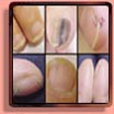
Nail Pitting:
Nail pitting -- small punctate depressions -- are caused by nail matrix inflammation, which can be the result of: (Figure 9); Figure 9: Indication of psoriasis. |
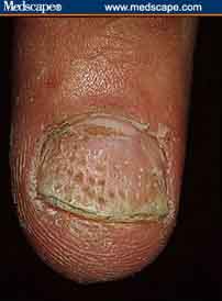
Figure 10: Alopecia areata. |
|
Nail Beading:
With nail beading, the beads seem to drip down the nail like wax (Figure 11). It is associated with endocrine conditions, including the following: |
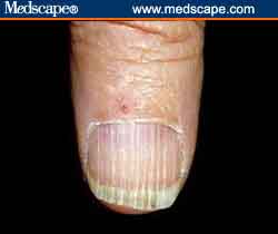
Figure 11: Nail beading. |
|
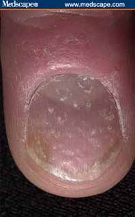
Rough Nail Surface:
When nails look sandpapered and dull, consider (Figure 12): Figure 12: Example of a rough nail surface. |
|
Nail Thickening:
Slow nail growth produces thickness (Figure 13). In such cases, the following should be considered: Onychomycosis, Chronic eczema, Peripheral vascular disease, Yellow nail syndrome & Psoriasis. Figure 13: Example of nail thickening. |
|
|
Onycholysis:
Onycholysis is distal separation of the nail plate from the underlying nail bed (Figure 14). It is associated with the following: Thyrotoxicosis, Psoriasis, Trauma, Contact dermatitis, Tetracycline, Eczema, Toxic exposures (solvents), Blistering from autoimmune disease, and Porphyria cutanea tarda (onycholysis and skin blistering from sun exposure). Figure 14: Traumatic onycholysis (involving only 1 nail). |
|
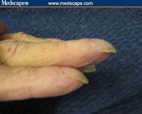
Severe Nail Curvature (Beaked Nails):
Curved or beaked nails are caused by resorption of distal digit (Figure 15). Consider the following: Hyperparathyroidism, Renal failure, Psoriasis & Systemic sclerosis Figure 15: Example of nail thickening. |
|
|
Complete Nail Destruction:
Complete local nail destruction can be caused by local mechanisms, including trauma and paronychia. Generalized conditions that might cause complete nail destruction include the following: |
CONTINUE THIS ARTICLE:
Previous Page - Page 2 of 5 - Next Page
MORE NAIL RESOURCES: