OBSERVING THE NAIL COLOUR
|
Abnormalities of the Lunula:
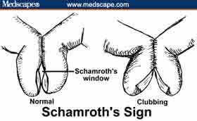
Nail clubbing involves a softening of the nail bed with the loss of normal Lovibond angle between the nail bed and the fold, an increase in the nail fold convexity, and a thickening of the end of the finger so it resembles a drumstick. To determine whether nails are clubbed, have the patient place both forefinger nails together and look between them. If you can see a small diamond space between them (Schamroth's window) then the nails are not clubbed (Schamroth's sign) (Figure 2). Causes of clubbing (not exhaustive) include the following (Figure 3): |
|
|
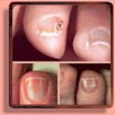
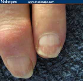
Figure 17: Pyramidal lunula. Figure 18: Lunula with red discoloration. |
|
|
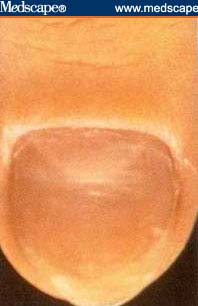
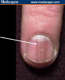
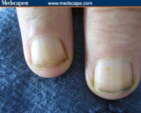
Transverse White Lines (Mee's lines)
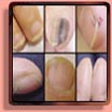
Any acute illness can produce transverse milky white lines. In addition, they might be caused by heavy metal toxicity (classically arsenic) or chemotherapy. The time of event may be determined from the location of the lines on nail (Figure 19). Figure 19: Note the Mee's line approximately one third of the way up the nail, suggesting a significant illness 2 months previously. |
|
|
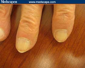
Leukonychia Striae
Leukonychia striata are white splotches caused by minor trauma to the nail matrix (Figure 20). The timing can be determined by the location of the splotches on the nail. Figure 20: Example of leukonychia striata. Note location of white splotches, which can indicate timing of the traumatic event. |
|
|
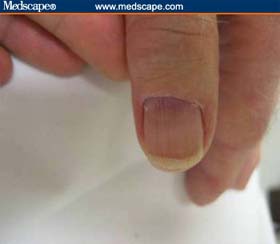
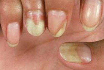
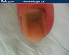
Longitudinal Brown Lines
Longitudinal brown lines form because of increased melanin produced by nail matrix melanocytes (Figure 21). They are associated with: Addison's disease; Nevus at the nail base; Breast cancer; Melanoma (check for periungal pigmentation) & Trauma. Figure 21: Longitudinal brown lines. |
|
|
Splinter Hemorrhages
Splinter hemorrhages are caused by hemorrhage of the distal capillary loop (Figure 22). Note the thickness of these areas. They are associated with the following: Subacute bacterial endocarditis; Systemic lupus erythematosus; Trichinosis; Pityriasis rubra pilaris; Psoriasis & Renal failure. Figure 22: Splinter hemorrhages tend to be fat. |
|
|
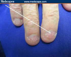
Terry's Half and Half Nails
With Terry's half and half nails, the proximal portion is white (edema and anemia) and the distal portion is dark. These nails imply either renal or liver disease (Figures 23). NOTICE: This piece of the article is not accurate, because Terry's nails and half-and-half nails involve two separate conditions with only a small overlap involving kidney problems. Figure 23: This example of Terry's half and half nails suggests liver disease (no brown lines). |
|
|
White Nails
White nails can be caused by anemia, edema, or vascular conditions (Figure 24). Consider the following: Anemia; Renal failure; Cirrhosis; Diabetes mellitus; Chemotherapy & Hereditary (rare). Figure 24: Example of white nails. |
|
|
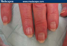
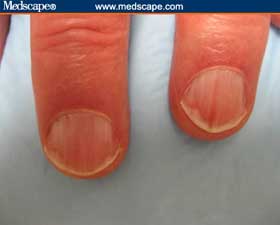
Pink or Red Nails
With pink or red nail discoloration, the following should be considered (Figure 25): Polycythemia (dark); Systemic lupus erythematosus; Carbon monoxide (cherry red); Angioma & Malnutrition. Figure 25: Example of pink and red nails. |
|
|
Brown-Gray Nails
Brown-gray nails may suggest the following (Figure 26): Cardiovascular disease; Diabetes mellitus; Vitamin B12 deficiency; Breast cancer; Malignant melanoma; Lichen planus; Syphilis & Topical agents, including hair dyes, solvents for false nails, varnish, and formaldehyde (among many others) Figure 26: Example of brown-gray nails. |
|
|
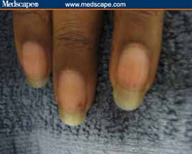
Yellow Nails
Yellow nails suggest the following (Figure 27): Diabetes mellitus; Amyloidosis; Median/ulnar nerve injury; Thermal injury & Jaundice. Consider yellow nail syndrome if a patient has lymphedema and bronchiectasis. Figure 27: Example of yellow nails. |
|
|
Green or Black Nails
Green or black nails indicate the following (Figure 28): Topical preparations, including chlorophyll derivations, methyl green, and silver nitrate (among others); Chronic Pseudomonas spp infection & Trauma. Figure 28: Example of black nails. |
|
CONTINUE THIS ARTICLE:
Previous Page - Page 3 of 5 - Next Page
MORE NAIL RESOURCES: